Proteomic Analysis of Ophthalmic Tissues

The vitreous is an optically clear, collagenous extracellular matrix that fills the inside of the eye and overlies the retina. Abnormal interactions between vitreous substructures and the retina underlie several vitreoretinal diseases, including retinal tear and detachment, macular pucker, macular hole, age-related macular degeneration, vitreomacular traction, proliferative vitreoretinopathy, proliferative diabetic retinopathy, and inherited vitreoretinopathies.
The molecular composition of the vitreous substructures is not known. Since the vitreous body is transparent with limited surgical access, it has been difficult to study its substructures at the molecular level. We developed a method to separate and preserve these tissues for proteomic analysis.
In other studies, we are collecting vitreous biopsies from patient eyes undergoing surgery. Analysis of the proteins in this fluid will reveal new biomarkers and therapeutic targets for eye disease.
Please contact us to learn how you can help support our research program.
News
Jun 16 2012 | Posted In: 20/20 Blog
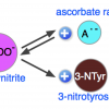
An NIH grant to study oxidative stress enzymes in the vitreous has important implications for treating diabetic vitreoretinopathy.

| Posted In: 20/20 Blog
The vitreous gel normally displays a graded density at different anatomical locations. Although it is estimated to be nearly 90% water, the vitreous gel contains diverse proteins, proteoglycans, and small molecules that originate from within and outside the eye. In specific vitreoretinal diseases,...